
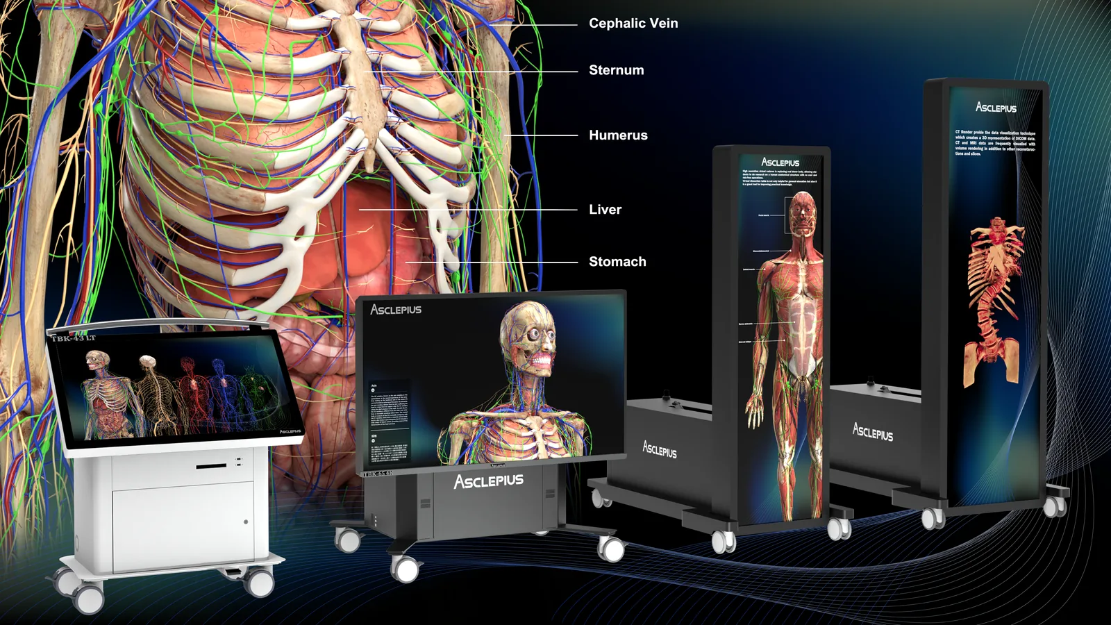
Medical Education
Say goodbye to cadaver shortages
The Asclepius Virtual Dissection Table puts a fully digitized human body on a touchscreen — over 5,000 anatomical structures that can be taken apart layer by layer, put back together, and dissected again tomorrow. No formalin, no odour, no waiting list.
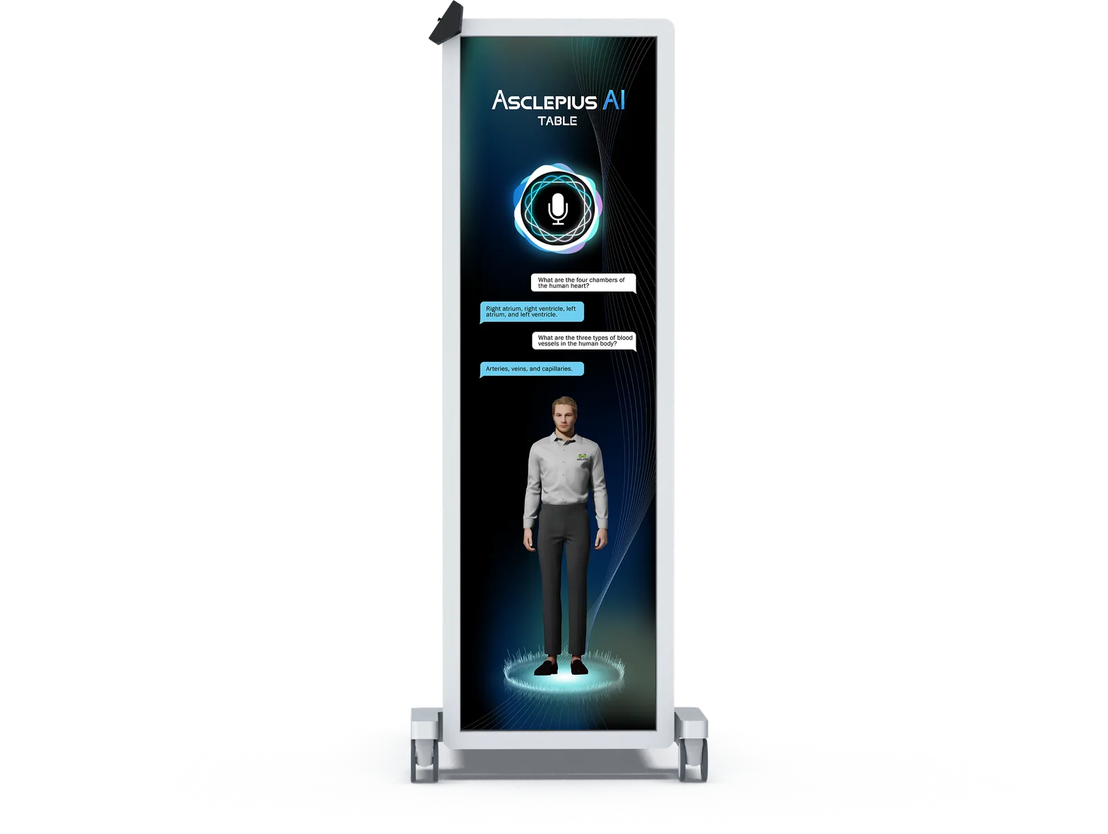
Asclepius AI Table
Asclepius AI Table
Intelligent labelling, content recommendations and a virtual assistant that listens. A built-in camera and microphone let the table recognize who is standing at it and answer questions out loud — so a class can ask about a structure instead of hunting for it.
- Facial recognition and voice tracking, with spoken commands and answers
- Eight software modules, adding Kinesiology and Ultrasound simulation
- Full-body physiological simulation for movement and systems analysis
- Exam questions from international nursing and medical licensure exams and the Taiwan registered nurse exams
- Menu reachable without moving, multilingual switching, side-by-side comparison
- Two models — TBK-AI-84 Ultra and TBK-AI-65 Vision 4K
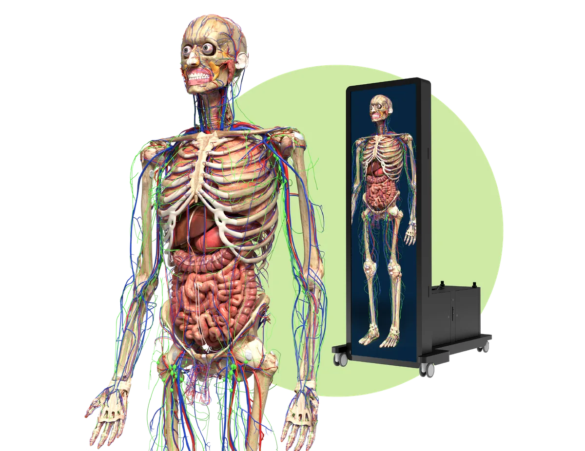
Virtual Dissection Table
One table, an entire anatomy department
Asclepius 3.0 is the standard virtual dissection table, in four sizes and without the AI assistant. Every structure is modeled and labeled, every dissection is reversible, and the content keeps growing — the result of years of collaboration between Surglasses, practising clinicians and anatomy professors.
- 5,000+ anatomical structures, dissectable layer by layer
- Anatomy teaching points and real cadaver images for comparison
- Clinical cases for teaching use only, plus your own DICOM uploads
- An exam question bank, with free online examination hosting
- Four sizes — 43″, 65″, 84″ and 99″ — from a lecture podium to a full anatomy lab
- Software updates included for the whole warranty period
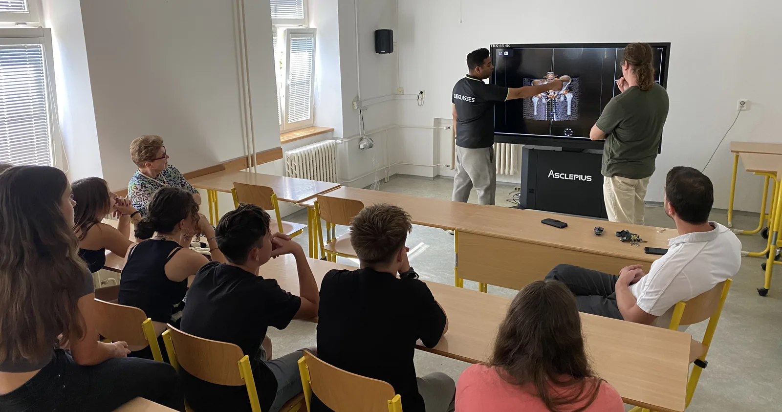
In the classroom
A different kind of anatomy lab
A digital table replaces or supplements traditional dissection in the teaching room.
No formalin, no odour
Nothing to preserve, ventilate or dispose of. The table removes the harmful chemicals, the smell and the maintenance bill that come with a traditional cadaver lab.
Unlimited dissections
A digital body can be cut apart as many times as the class needs and restored instantly. Teaching is no longer rationed by how many specimens the department managed to obtain this year.
Built for groups
Two to four people around the 43″, seven to ten around the 84″ and 99″. Everyone sees the same body at the same time, so the discussion happens over the model rather than over a textbook.
What’s inside
Six software packages, one interface
All six packages are standard on the 65, 84 and 99-inch tables; the 43-inch lecture table includes Anatomy and OOOPDS Radiology as standard. The AI Table carries all six plus Kinesiology and Ultrasound simulation.
Anatomy
Full-body cadaver models covering both sexes and several ethnicities, teaching points, real cadaver images for comparison, and five interactive endoscopic simulations.
OOOPDS Radiology
Radiology module based on OOOPDS (FDA 510(k) K254214), for educational purposes only. Clinical cases for teaching use, semi-automatic bone segmentation, and conversion of 2D scans into 3D models that can be sent straight to a 3D printer.
Histopathology Atlas
Digitized histology and pathology slides, captured so they stay sharp all the way down to the cell, with expert annotations.
CT Render
Renders DICOM data into interactive 3D volumes, separating soft tissue from bone with adjustable transparency, contrast and color, and slicing freely in any anatomical plane.
Embryology
Week-by-week fetal development from week 5 to week 42, Carnegie stages, the anatomy of pregnancy with maternal–fetal blood exchange, and over 100 documented embryonic pathologies.
Animal Anatomy
Animal species modeled in 3D for comparative anatomy, each structure clickable for its name and function, and dissectable layer by layer like the human models.
Every table also comes with the Online Examination platform — teachers set the topic, difficulty and time limit, and grading is instant — and the Asclepius Anatomy Online app for Windows, so students can keep revising away from the table.
Explore
Who uses it, and what for
The same table serves a high-school biology class and a hospital simulation center — what changes is the software, the size and the lesson plan.
Institutions
Medical simulation centers, medical schools, undergraduate healthcare programs and K–12 science classrooms — four very different audiences, each with its own configuration and curriculum fit.
See the four segments
Applications
Cadaver labs, clinical training, surgical planning practice and patient communication.
See how it is usedSee it for yourself
Find the table that fits your teaching room
Tell us the size of your classes and the courses you run, and we will show you the model and software combination that makes sense — online, or on site with the table in front of you.